-40%
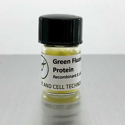
Green Fluorescent Protein
$ 26.4
- Description
- Size Guide
Description
GREEN FLUORESCENT PROTEINThe Green Fluorescent Protein (GFP) from A. Victoria has revolutionized molecular biology. Applications such as GFP tagging and GFP reporter genes have allowed scientists to image the activity of proteins in living cells and even entire organisms.
Here we provide recombinant GFP protein, produced in E. coli. It may be used as a control for experiments involving GFP tags, or for any research purpose the creative mind can imagine.
Like all our products,
GFP
is sold for research use only. It is not for human use.
GFP BIOCHEMISTRY
The amino-acid sequence of GACT recombinant GFP is:
GVSKGEELFTGVVPILVELDGDVNGHKFSVSGEGEGDATYGKLTLKFICTTGKLPVPWPTLVTTL TYGVQCFSRYPDHMKQHDFFKSAMPEGYVQERTIFFKDDGNYKTRAEVKFEGDTLVNRIELKGIDFKE DGNILGHKLEYNYNSHNVYIMADKQKNGIKVNFKIRHNIEDGSVQLADHYQQNTPIGDGPVLLPDNHYLSTQSALSKDPNEKRDHMVLLEFVTAAGITLGMDELYK
Mw = 26.9 kDa
pI = 5.59
e280 = 21.9 mM-1 cm-1
e488 = 41.8 mM-1 cm-1
Compared with natural GFP, this GFP has mutations F64L and S65T. This has been reported to dramatically enhance its brightness (PMID 8707053). Others have given the name enhanced GFP, or "eGFP" to this sequence.
SPECTRA
The characteristic absorbance maxima at 280 (aromatic amino acids) and 488 (GFP chromophore) are visible. The ratio of the two can predict the purity of the GFP and the degree of successful chromophore formation. Our ratio is guaranteed >1.5, and is usually above 1.9.
GFP can be excited either at its blue light maximum 488 nm, or very effectively in the UV range 280 - 300 nm. UV exposure however will photobleach the GFP quickly. It is quite remarkably resistant to photobleaching at 488 nm. The emission maximum is at 510 nm.
Successful fluorescence experiments generally depend on the use of filters that can fully exclude the exciting beam from the detector that measures the emitted wavelength. Most generally available fluorescence lab equipment will come with "GFP" labeled filters that accomplish this well. If that's for some reason not the case, it is certainly possible to choose suboptimal wavelengths based on these spectra, with a corresponding loss of brightness intensity.
STORAGE & HANDLING
Our lyophilized GFP cake is highly porous, voluminous and light. It is stable at ambient temperature during shipping. Upon receipt, it can be stored at +4C, -20C or -80C for years. It is essential to keep moisture away from the lyophilisate. Let the vial come to room temperature before opening to prevent condensation of water from the air onto the lyophilisate. In this way, solid amounts can be removed from the vial with a spatula repeatedly.
Alternatively, you may dissolve all the GFP in the vial in water or a biological buffer of your choice. Adding a small amount of water will draw the entire cake into solution quickly. Once dissolved, GFP should be stored frozen at -80C. GFP is resistant to many freeze-thaw cycles, but may degrade if stored liquid in the fridge (within weeks).
GFP is also surprisingly resistant to light exposure. It will however be damaged if exposed to bright light for a long period of time. The product ships in a small cardboard box that is ideal to block any incident light out during storage. The vial itself is made out of light transmissive glass.
PURITY
The purity of our GFP is assessed by SDS-PAGE with Coomassie staining:
No contaminant bands must be detected with 2 ug total protein loading volume and sensitive Coomassie G-250 staining / destaining protocol. As an additional measure of purity the ratio of absorbance A488 / A280 must be >1.5.
COMPOSITION
Prior to lyophilization, we bring GFP to 5 mg/ml in 10 mM Tris pH 8.2. The Tris is still present in the lyophilizate. We prepare our tris buffer using Hasselbalch's equation from Tris-OH and Tris-HCl. There is no after-the-fact pH adjustment. The amount of buffer salts in the lyophilizate are thus predictable for those who need to know. There are no other additives.
The GFP amount present in the vial is determined using an ideal GFP extinction coefficient of e488 = 55 mM-1 cm-1. As always, we try to give you a little bit more than advertised. If your experiment requires precise titration, it is best to do your own spectrophotometric determination.


